science has advanced exponentially in the last century. Even just the way the human body and pathology are viewed. We’ve gone from the basic x-ray to be able to see a single cell in the human body. Or even smaller.
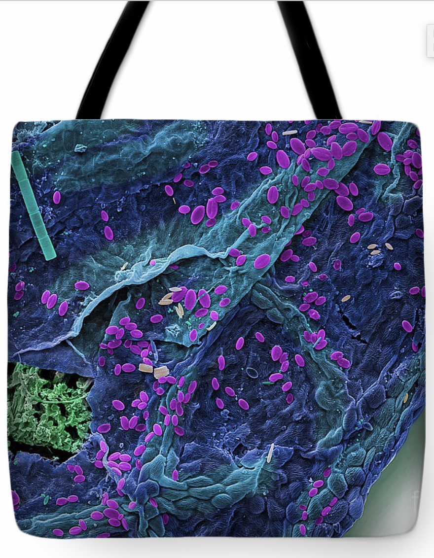
One of the most fascinating innovations has been the Scanning Electron Micrograph, often referred to simply as an SEM for short. It has revealed hidden worlds of the human body, medical, and of course the natural world as well.
Gallery of Medical and Anatomical SEMs (micrographs) Stock Photography
The scanning electron microscope was invented in 1937 by Manfred von Ardenne the SEM machine uses electrons to record the surface topography of objects.