Pages

Showing posts with label microscope. Show all posts

Sunday, November 28, 2021

Microscope Photography (SEMS) Make Unique Gifts for the Science Minded


Woman uses a Scanning Electron Microscope (left) 
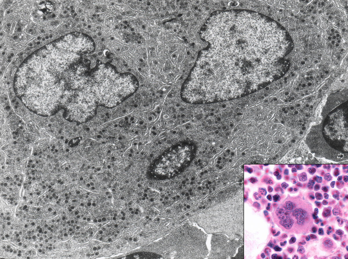
, a cell heavily infected withARS-CoV-2 virus particles (right).

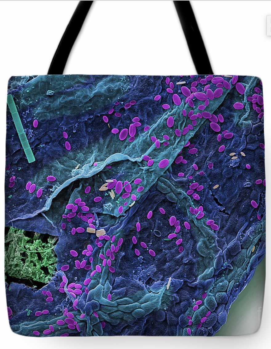
 The entire world has become more familiar with micrographs in the past 18 months, although you may not have realized it. During the pandemic, detailed photographs of the coronavirus appeared in newspapers, television, and everywhere online.  They were taken by a particular device called a scanning electron microscope. The images are often referred to as SEMs for short.

As the name implies, the microscope uses a particle beam to detect electrons off the surface of specimens, which is then placed in a vacuum to create sharp images. Magnification ranges from 20x to 30,000x, spatial resolution of 50 -100nm.

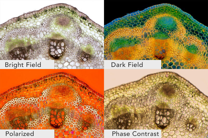
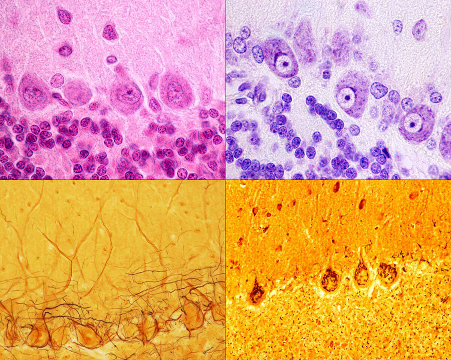
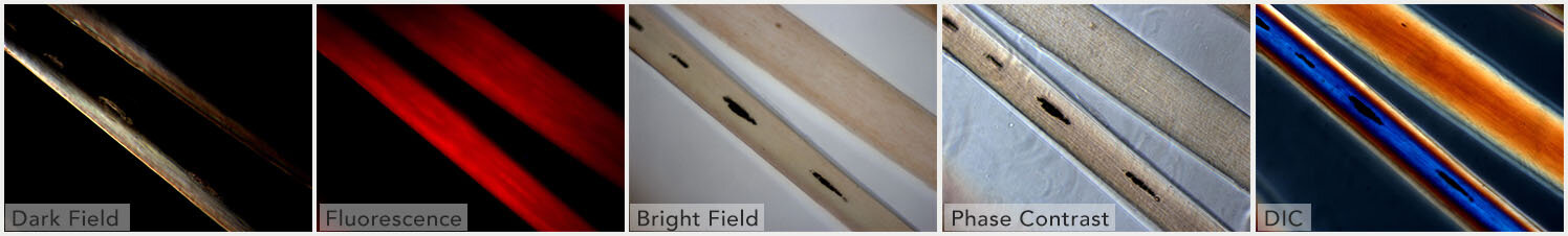
Micrographs allow us to see critical details that may be useful in curing diseases, and they shows us the hidden beauty of everyday objects.


A fine art print, a fun t-shirt, mug, or jigsaw puzzle of a micrograph will make a bold impression on anyone. Resembling abstract art, they’re great for people in the sciences, medicine, research, or just someone who enjoys nature. Discover what micrograph gifts you can make!


Peruse these fascinating images at our Fine Art America shop to start creating amazing micrograph gifts!


Tuesday, January 5, 2021

Taking a Closer Look at Microscopy for Medical and Scientific Use

Waterbear Micrograph

 Read any scientific or medical news story and you can see that microscopy has come a long way since your high school biology class microscope.

It isn't just higher magnification, but crisp details, a greater depth of field, viewing internal features, and colorful 3D-like visuals that fascinate us. There have been many advancements to light microscopes and a multitude of new kinds that can see so much more than we could have ever expected.  

Stock Micrographs

Let's take a closer look!

Four types of Light micrographs: Bright Field, Dark Field, Polarized, Phase Contrast. © Marek Mis/Science Source

Four types of Light micrographs: Bright Field, Dark Field, Polarized, Phase Contrast. © Marek Mis/Science Source

The basic microscope we used as a kid is the standard “light microscope”. Simply put, it uses light and a set of lenses. The addition of filters, specialized mirrors, lasers, specific light spectrums, and other features gives us much more detail.

More advanced devices include Scanning Electron Microscopes (SEM), Transmission Electron Microscopes (TEM), Atomic Force Microscopes (AFM), and Scanning Tunneling Microscopes (STM).

Commonly used techniques when viewing slides on a light microscope are Bright Field, Dark Field, Fluorescence, Differential Interference Contrast (DIC), Phase Contrast, and Confocal microscopy.

Light MIcroscope Bright Field: the light source shines directly from underneath the specimen, creating a light-colored or bright area around it.

Purkinje neurons of the cerebellar cortex stained with four staining methods: hematoxylin eosin (top left), cresyl violet (top right), Cajal's silver nitrate (bottom left) and silver method for Golgi apparatus (bottom right). © Jose Luis Calvo/Science Source

Purkinje neurons of the cerebellar cortex stained with four staining methods: hematoxylin eosin (top left), cresyl violet (top right), Cajal's silver nitrate (bottom left) and silver method for Golgi apparatus (bottom right). © Jose Luis Calvo/Science Source

Light Microscope Dark Field: the light source is occulted, so it reaches the specimen at different angles giving us slightly more varied details than if it was lit from directly underneath. The area around the specimen is dark or black.

Fluorescence: This uses light filters and specific wavelengths. Short wavelengths are reflected down to the specimen, which then fluoresces or gives off long wavelengths of light. These are reflected up to a mirror that allows long wavelengths to pass through to the lens.

Phase Contrast: Using a special lens and filters it allows viewing of transparent and colorless specimens. It looks similar to DIC micrography but lacks shadows, making it a bit more two dimensional.

Different illumination techniques of a light microscope: dark field, fluorescence, bright field, phase contrast, DIC (differential interference contrast). Child’s hair strand © Ted Kinsman/Science Source

Different illumination techniques of a light microscope: dark field, fluorescence, bright field, phase contrast, DIC (differential interference contrast). Child’s hair strand © Ted Kinsman/Science Source

Differential Interference Contrast Microscopy (DIC): Using a  polarizer, beam splitter, condenser, and filters it allows viewing of transparent and colorless specimens. It has a more three-dimensional appearance than phase-contrast microscopy.

Confocal Microscopy: Also called Confocal Laser Scanning Microscopy (CLSM), it uses a laser and a spatial pinhole to create a sharper image.

Let's look at the more advanced types of microscopes:

|Atomic Force Micrograph of plasma membrane proteins.

|Atomic Force Micrograph of plasma membrane proteins.

Scanning Electron Microscope SEM: Uses a particle beam of electrons. It detects reflected electrons off the surface of a specimen, which is placed in a vacuum. creating sharp images. Magnification ranges from 20x to 30,000x, spatial resolution of 50 - 100nm.

Bone marrow cell. This micrograph shows this cell with light (inset) and electron microscope. © Jose Luis Calvo/Science Source

Bone marrow cell. This micrograph shows this cell with light (inset) and electron microscope. © Jose Luis Calvo/Science Source

Transmission Electron Microscope TEM: Uses a particle beam of electrons that pass through a thinly sliced specimen. It can show the internal structures of cells with a magnification up to 2,000,000x.

Atomic Force Microscope AFM: AFM uses a laser that bounces off of a stylus on a cantilever lever. This action traces the specimen. Any deviation triggers the sensors creating a raster image. One benefit of this is that it also records the Z-Plane. Another advantage of AFM over electron microscopy is that the specimen need not be in a vacuum.

Scanning Tunneling Microscope STM: Scanning Tunneling Microscope STM: An STM also uses electrons, based on quantum tunneling. The benefits are that it can be used in a vacuum, air, water, or ambient gas environment. It captures surfaces on the atomic level.

What can we look at with all of these scopes? 

Scoop up pond water or ocean water to be astonished by the plethora of living zooplankton and phytoplankton visible within a single drop using a simple light microscope. 

It opens you to the wonder of cyanobacteria,  blue-green algae, ciliates like paramecium, daphnia, amoebas, and euglena. If you were lucky, you might have witnessed them conjugate and divide!

Additionally,  the ocean water drop allows a peek at copepods, immature mollusks, krill, algae, crustaceans, fish in their zooplankton stage; and you may behold the breathtaking beauty of diatoms, the most common type of phytoplankton in our oceans. 

Switch to a higher-powered Scanning Electron Microscope (SEM) to view Water Bears, pollen, blood cells, and insects. An SEM uses a particle beam of electrons to photograph the surface of a vacuum-sealed specimen.

Transmission Electron Microscopes(TEM) allows us to see cross-sections of a specimen like the beautiful interior of the human body, marine life, and animal and plant cells. The TEM's particle beam passes through its vacuum sealed specimen. 

micrograph-shower-curtain.png

Custom homedecor, phone cases, shirts and more.

Of course, there are critical medical applications. 

We can view the cells of the human body with many different microscopes. The light mic, SEM, and TEM show scientists and medical researchers different angles and aspects of the cell and its fine structures and organelles. 

Microscopes help scientists study cancer - breast, ovarian, prostate, liver, and skin cancer.  We can develop an improved understanding of skin conditions such as psoriasis and eczema. They assist in the fight against nervous, respiratory, and circulatory system diseases. And a cure for muscular conditions such as fibromyalgia and multiple sclerosis (MS) and autoimmune conditions.

Infectious agents such as bacteria, viruses, fungi, prions, and parasites can be examined. It allows us to better understand, diagnose, and work towards cures, vaccines, or prevention. 

And without the microscope, how could we progress in the fight against the seasonal flu, measles, polio, malaria, and HIV/AIDS.

If light, lasers, electrons, and quantum physics are not enough, there is even a microscopy method, similar to SONAR, that uses sound waves; Acoustic Microscopy.

Lastly, without these microscopes, we would not be able to continue our current fight against the Coronavirus, COVID-19.


Tuesday, December 22, 2020

Medical Micrographs: a View into the Human Body


science has advanced exponentially in the last century. Even just the way the human body and pathology are viewed. We’ve gone from the basic x-ray to be able to see a single cell in the human body. Or even smaller.

One of the most fascinating innovations has been the Scanning Electron Micrograph, often referred to simply as an SEM for short. It has revealed hidden worlds of the human body, medical, and of course the natural world as well.

Gallery of Medical and Anatomical SEMs (micrographs) Stock Photography

The scanning electron microscope was invented in 1937 by Manfred von Ardenne the SEM machine uses electrons to record the surface topography of objects.

SEM of Maple Leaf Tote bag

SEM of Maple Leaf Tote bag

Sunday, October 14, 2018

Explore the Human Microbiome


The idea of bacteria creeping through your body might be less than appetizing. We often associate the presence of bacteria in our bodies (especially in our digestive system) with infections, viruses, and food poisonings. However, scientific findings have uncovered the fact that there are also many types of good bacteria that are essential to our health. This collection of good and bad bacteria within our bodies have also been discovered to be strikingly diverse from person to person.

The definition of the human microbiome is still somewhat ambiguous and disputed but is commonly known to be the vast ecosystem of microscopic organisms (microbes) living within us. According to the National Center for Biotechnology Information (NCBI), we consist of 10-100 trillion microbiota, also known as microbial cells. These cells hold genes that determine cell behavior.

Stock Images and Stock Video of Microbes


Scientists have become specifically interested in the gut (our stomach and intestines) as a location that holds a notably vast collection of microbes. Believe it or not, the large intestine houses good bacteria, such as probiotics, that fight the bad bacteria and keeps us healthy. The ratio of good bacteria to bad can change and partly depends on your immune system or diet choices. If good bacteria becomes low in numbers, risk of infection and virus development increases.

One of the fascinating aspects of our microbiomes is that everyone's is distinctly unique. We, as humans, have a much different microbiome than other animals, but ours also differ from person to person. Even the collection of organisms in your mouth is different than the community of organisms in your gut.

Because each person's microbiome is so individualized, researchers are recognizing that this information could be valuable for practical usage in medicine. A patient's microbiome could be used similarly to a fingerprint, but instead of merely identifying the patient, medical professionals could gain a better understanding about what is going on within the patient's body and be able to make more educated predictions about procedures and treatments needed.

Microbial Mugs, Notebooks, and Other Custom Gifts


As more and more scientific findings of this topic surface, it becomes increasingly more exciting to find out how this knowledge will help us solve many unanswered questions that have to do with anatomical behavior, virus prevention, and even evolution. Our microbiomes seem to be the mysterious key that could open many doors to the understanding of the human body.


  • learn.genetics.utah.edu/content/microbiome
  • https://www.ncbi.nlm.nih.gov/pmc/articles/PMC3426293
  • http://www.nutritionnews.abbott/nutrition-as-medicine/the-role-of-the-microbiome-in-gut-health-

Rotifers, Nematodes and Tardigrades Stock Microscopic Photography

 Roti fers (Philodina sp.), Light Micrograph The bdelloid rotifer, found in freshwater habitats all over the world, is able to withstand ex...