Pages

Showing posts with label science. Show all posts

Friday, September 8, 2023

Rotifers, Nematodes and Tardigrades Stock Microscopic Photography


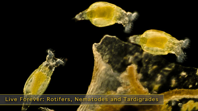
 Rotifers (Philodina sp.), Light Micrograph


The bdelloid rotifer, found in freshwater habitats all over the world, is able to withstand extreme cold. According to a recent discovery in northeastern Siberia, these multicellular organisms can be frozen for up to 24,000 years and live to tell the tale!

The average life span of us humans, 78 years, is not much compared to other creatures. Some birds live up to 100 years, eels have been recorded at 106, tortoises 150, and Koi fish over 200. The slow moving Greenland shark has been recorded at 512 years.

Still these are just flashes in the pan compared to rotifers. Scientists recently restored rotifers that had been frozen in the Siberian permafrost for over 24,000 years, meaning these creatures were alive during the Late Pleistocene Era - when Wooly Mammoths roamed the earth.

Stock Images and Video of Rotifers and Other Long-lived Animals

A rotifer is part of a group of seemingly invincible creatures, such as nematodes and tardigrades. Tardigrades have even been sent to outer space and survived.

Rotifers are a type of microscopic animal that is often found in zooplankton in either freshwater or saltwater. Rev. John Harris, in 1696, was the first to mention these creatures. They are commonly referred to as wheel animals due to the motion of the cilia surrounding their mouths, which looks like a spinning wheel.

Rotifers are filter feeders that eat dead bacteria and other decaying organic matter. When they move around, they compress their bodies into round shapes and extend out into a long thin shape. In terms of size they can range from 50 micrometers to over 2 millimeters.


Sunday, November 28, 2021

Microscope Photography (SEMS) Make Unique Gifts for the Science Minded


Woman uses a Scanning Electron Microscope (left) 
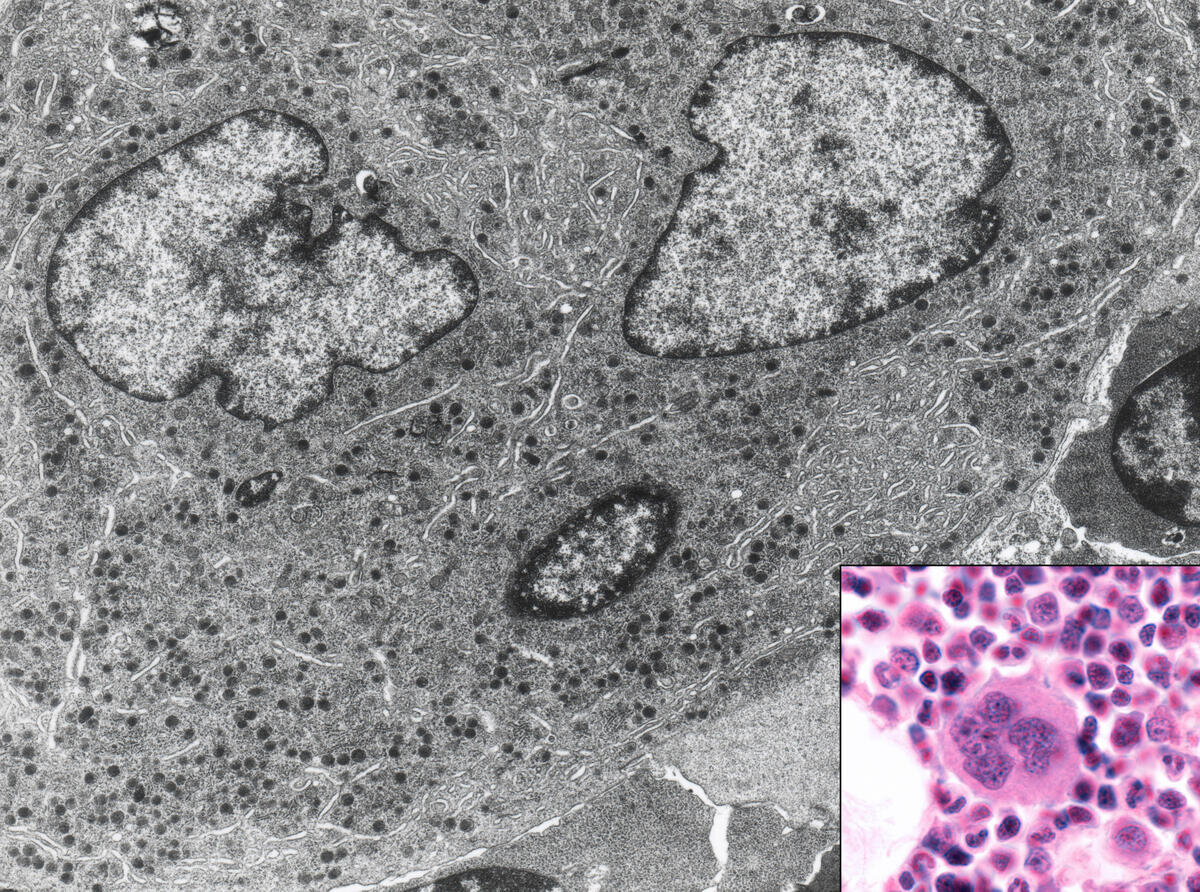
, a cell heavily infected withARS-CoV-2 virus particles (right).

 The entire world has become more familiar with micrographs in the past 18 months, although you may not have realized it. During the pandemic, detailed photographs of the coronavirus appeared in newspapers, television, and everywhere online.  They were taken by a particular device called a scanning electron microscope. The images are often referred to as SEMs for short.

As the name implies, the microscope uses a particle beam to detect electrons off the surface of specimens, which is then placed in a vacuum to create sharp images. Magnification ranges from 20x to 30,000x, spatial resolution of 50 -100nm.

Micrographs allow us to see critical details that may be useful in curing diseases, and they shows us the hidden beauty of everyday objects.


A fine art print, a fun t-shirt, mug, or jigsaw puzzle of a micrograph will make a bold impression on anyone. Resembling abstract art, they’re great for people in the sciences, medicine, research, or just someone who enjoys nature. Discover what micrograph gifts you can make!


Peruse these fascinating images at our Fine Art America shop to start creating amazing micrograph gifts!


Monday, February 1, 2021

Super Earths and Exoplanets

Super Earths


we explore further afield from our home planet, we learn there are not only other systems, but many other planets in these systems. There are people who look to these for another earth and for alien life.

Planets outside of our solar system are called exoplanets.

Gallery of Stock Pictures of Super-Earths and Exoplanets

Just as our solar system has planets of different sizes and types, planets in other systems are varied in size and composition.

To date, we have found 3,242 systems with planets, many with multiple planets, bringing the number of exoplanets we know of to 4,395.

Planets are categorized by their mass. Any planet with a mass larger than earth without being 14 times higher in mass is considered a super-earth. 

The designation doesn't speak to its composition or whether it would be a friendly place for us humans to live. It only tells us the comparative mass of an exoplanet.

Recently, NASA discovered a new super-planet naming it TOI-561b.

TOI-561b travels around its sun twice as fast as we travel around ours. So it experiences two days in the time we experience one.

Life is unlikely to exist there as the temperature on it is 3,140 degrees Fahrenheit.

Yet, it is exciting to keep discovering more and more systems and planets. Perhaps, one day, we'll find one more compatible with life on our earth,

Thursday, January 28, 2021

The Importance of Protein Folding


Tuesday, January 5, 2021

Taking a Closer Look at Microscopy for Medical and Scientific Use

Waterbear Micrograph

 Read any scientific or medical news story and you can see that microscopy has come a long way since your high school biology class microscope.

It isn't just higher magnification, but crisp details, a greater depth of field, viewing internal features, and colorful 3D-like visuals that fascinate us. There have been many advancements to light microscopes and a multitude of new kinds that can see so much more than we could have ever expected.  

Stock Micrographs

Let's take a closer look!

Four types of Light micrographs: Bright Field, Dark Field, Polarized, Phase Contrast. © Marek Mis/Science Source

Four types of Light micrographs: Bright Field, Dark Field, Polarized, Phase Contrast. © Marek Mis/Science Source

The basic microscope we used as a kid is the standard “light microscope”. Simply put, it uses light and a set of lenses. The addition of filters, specialized mirrors, lasers, specific light spectrums, and other features gives us much more detail.

More advanced devices include Scanning Electron Microscopes (SEM), Transmission Electron Microscopes (TEM), Atomic Force Microscopes (AFM), and Scanning Tunneling Microscopes (STM).

Commonly used techniques when viewing slides on a light microscope are Bright Field, Dark Field, Fluorescence, Differential Interference Contrast (DIC), Phase Contrast, and Confocal microscopy.

Light MIcroscope Bright Field: the light source shines directly from underneath the specimen, creating a light-colored or bright area around it.

Purkinje neurons of the cerebellar cortex stained with four staining methods: hematoxylin eosin (top left), cresyl violet (top right), Cajal's silver nitrate (bottom left) and silver method for Golgi apparatus (bottom right). © Jose Luis Calvo/Science Source

Purkinje neurons of the cerebellar cortex stained with four staining methods: hematoxylin eosin (top left), cresyl violet (top right), Cajal's silver nitrate (bottom left) and silver method for Golgi apparatus (bottom right). © Jose Luis Calvo/Science Source

Light Microscope Dark Field: the light source is occulted, so it reaches the specimen at different angles giving us slightly more varied details than if it was lit from directly underneath. The area around the specimen is dark or black.

Fluorescence: This uses light filters and specific wavelengths. Short wavelengths are reflected down to the specimen, which then fluoresces or gives off long wavelengths of light. These are reflected up to a mirror that allows long wavelengths to pass through to the lens.

Phase Contrast: Using a special lens and filters it allows viewing of transparent and colorless specimens. It looks similar to DIC micrography but lacks shadows, making it a bit more two dimensional.

Different illumination techniques of a light microscope: dark field, fluorescence, bright field, phase contrast, DIC (differential interference contrast). Child’s hair strand © Ted Kinsman/Science Source

Different illumination techniques of a light microscope: dark field, fluorescence, bright field, phase contrast, DIC (differential interference contrast). Child’s hair strand © Ted Kinsman/Science Source

Differential Interference Contrast Microscopy (DIC): Using a  polarizer, beam splitter, condenser, and filters it allows viewing of transparent and colorless specimens. It has a more three-dimensional appearance than phase-contrast microscopy.

Confocal Microscopy: Also called Confocal Laser Scanning Microscopy (CLSM), it uses a laser and a spatial pinhole to create a sharper image.

Let's look at the more advanced types of microscopes:

|Atomic Force Micrograph of plasma membrane proteins.

|Atomic Force Micrograph of plasma membrane proteins.

Scanning Electron Microscope SEM: Uses a particle beam of electrons. It detects reflected electrons off the surface of a specimen, which is placed in a vacuum. creating sharp images. Magnification ranges from 20x to 30,000x, spatial resolution of 50 - 100nm.

Bone marrow cell. This micrograph shows this cell with light (inset) and electron microscope. © Jose Luis Calvo/Science Source

Bone marrow cell. This micrograph shows this cell with light (inset) and electron microscope. © Jose Luis Calvo/Science Source

Transmission Electron Microscope TEM: Uses a particle beam of electrons that pass through a thinly sliced specimen. It can show the internal structures of cells with a magnification up to 2,000,000x.

Atomic Force Microscope AFM: AFM uses a laser that bounces off of a stylus on a cantilever lever. This action traces the specimen. Any deviation triggers the sensors creating a raster image. One benefit of this is that it also records the Z-Plane. Another advantage of AFM over electron microscopy is that the specimen need not be in a vacuum.

Scanning Tunneling Microscope STM: Scanning Tunneling Microscope STM: An STM also uses electrons, based on quantum tunneling. The benefits are that it can be used in a vacuum, air, water, or ambient gas environment. It captures surfaces on the atomic level.

What can we look at with all of these scopes? 

Scoop up pond water or ocean water to be astonished by the plethora of living zooplankton and phytoplankton visible within a single drop using a simple light microscope. 

It opens you to the wonder of cyanobacteria,  blue-green algae, ciliates like paramecium, daphnia, amoebas, and euglena. If you were lucky, you might have witnessed them conjugate and divide!

Additionally,  the ocean water drop allows a peek at copepods, immature mollusks, krill, algae, crustaceans, fish in their zooplankton stage; and you may behold the breathtaking beauty of diatoms, the most common type of phytoplankton in our oceans. 

Switch to a higher-powered Scanning Electron Microscope (SEM) to view Water Bears, pollen, blood cells, and insects. An SEM uses a particle beam of electrons to photograph the surface of a vacuum-sealed specimen.

Transmission Electron Microscopes(TEM) allows us to see cross-sections of a specimen like the beautiful interior of the human body, marine life, and animal and plant cells. The TEM's particle beam passes through its vacuum sealed specimen. 

micrograph-shower-curtain.png

Custom homedecor, phone cases, shirts and more.

Of course, there are critical medical applications. 

We can view the cells of the human body with many different microscopes. The light mic, SEM, and TEM show scientists and medical researchers different angles and aspects of the cell and its fine structures and organelles. 

Microscopes help scientists study cancer - breast, ovarian, prostate, liver, and skin cancer.  We can develop an improved understanding of skin conditions such as psoriasis and eczema. They assist in the fight against nervous, respiratory, and circulatory system diseases. And a cure for muscular conditions such as fibromyalgia and multiple sclerosis (MS) and autoimmune conditions.

Infectious agents such as bacteria, viruses, fungi, prions, and parasites can be examined. It allows us to better understand, diagnose, and work towards cures, vaccines, or prevention. 

And without the microscope, how could we progress in the fight against the seasonal flu, measles, polio, malaria, and HIV/AIDS.

If light, lasers, electrons, and quantum physics are not enough, there is even a microscopy method, similar to SONAR, that uses sound waves; Acoustic Microscopy.

Lastly, without these microscopes, we would not be able to continue our current fight against the Coronavirus, COVID-19.


Friday, November 1, 2019

Rising Sea Levels

One of the most significant challenges related to global climate change is rising sea levels. Since the beginning of the 20th-century, sea levels have gradually risen by 16 - 21 cm (6.3 - 8.3 in).

The rise in sea levels has also accelerated over time due to thermal expansion (the change in the volume and area of matter due to increased temperatures) and the melting of ice sheets and arctic glaciers.


Researchers have calculated that arctic sea ice has declined by 10% in the last 10 years. In addition to rising sea levels, this has altered the circulation of the Atlantic Ocean, destroying fisheries in northern countries and creating massive storms and hurricanes.

Coastlines around the world remain susceptible to flooding. Some at-risk coasts include the Miami shore, Rio de Janeiro, Osaka and Shanghai. The Egyptian city, Alexandria, faces a particular threat as much of its population exists in low lying coastal areas, which will likely be inundated within the next decade.


Science phone cases, mugs and more

Science phone cases, mugs and more
Our best hope for limiting the increase in sea levels is to reduce our carbon footprint. Initiatives in renewable energy and efficient power are critical components in reaching our emission goals.
For images of climate science and rising sea levels, click the links above. For information on how you can help deduce our carbon emissions head to the Environmental Defense Fund. (www.edf.org)

Tuesday, July 30, 2019

Immunotherapy: Your Immune System is Cancer's Biggest Enemy


There’s no doubt about it - our immune systems are powerful. We’ve all had moments where we don’t pay much attention to our immune systems because of lack of sleep or high stress and had to endure bouts of sneezing, coughing, or fever in the coming days. Some of us have taken extra steps to make sure our immune systems are as strong as possible and noticed how invincible it feels to go months without getting sick. Everyday function, brain power, and physical ability depend strongly on our body’s ability to fight off viruses and disease.

The power that our immune system provides in determining our health can even be utilized to fight cancer. Immunotherapy is a method of cancer treatment that helps to strengthen the immune system to be able to fight cancer cells better, as well as direct the immune system towards specific cancer cells.

RF and RM Images and Video of Immunotherapy

A large number of cells in our body aid in the immune system. White blood cells (lymphocytes) recognize foreign bodies such as viruses and fight them with antibodies, which are proteins that are sent out to bind to these foreign bodies. T cells are a specific type of white blood cell that search for, capture and destroys foreign bodies and infected cells.

Immunotherapy works with these cell types to focus in on cancer cells and kill as many as possible. There are many different methods of immunotherapy that enhance the immune system in a variety of ways.

Proteins called monoclonal antibodies (therapeutic antibodies) can be created in a laboratory and used for immunotherapy. These antibodies are introduced into the body to flag cancer cells so the immune system can more easily detect them.

Checkpoint inhibitors can be put into a patient’s bloodstream to disable proteins that stop the immune system from attacking cancer cells. These proteins attach to T cells, and can activate or deactivate the immune system when needed. Because cancer cells can pose as normal cells and pass a T cell checkpoint without any activation of the immune system, checkpoint inhibitors are used to make sure that T cells will be activated to help destroy cancer cells with each encounter.

Adoptive cell transfer is a form of therapy that can also help boost the function of T cells. In this method, T cells are taken from the body, and the cells that are most defensive are grown and multiplied in a lab and put back into the body intravenously.

Prints of Cancer and the Immune System

Through the technology that scientists now have available in labs, immunotherapy provides another option for cancer treatment which can give many patients more hope. Although this type of therapy has its own side effects, and sometimes has the potential to damage the body, it has been known to successfully treat some cancers when radiation or chemotherapy failed, and can also enhance the effectiveness of other methods of treatment when used in combination.

Overall, it has the potential to provide hope to many of millions of people around the world affected by cancer.

  • livescience.com
  • cancer.gov
  • immunology.org
  • pacificimmunology.com
  • tcells.org

Tuesday, July 23, 2019

Origins of Modern Chemistry


How did we get from discovering fire to using nanotubes in labs?

The concept of atoms was first conceived in ancient Greece by a group of philosophers known as the atomists. In 330 BC, Aristotle opposed this theory. He was a proponent of the elemental theory, positing that all matter was composed of the elements earth, fire, water, and air. He even added an element, aether (or ether).

The elemental theory continued into the middle ages through the study of alchemy. Alchemists added sulfur, salt, and mercury to the list.

RF and RM Stock Images and Video
of the History of Modern Chemistry

Finally, in 1661, Robert Boyle published the book "The Sceptical Chymist", presenting his hypothesis of particles in motion and asserting that only experiments using the "scientific method" could be considered true.

Another milestone in chemistry was the discovery of electrochemistry. Alessandro Volta invented the first battery in 1800, using piles of copper and zinc discs. They were separated by cloth soaked in brine. When wires were attached to the top and bottom, electricity flowed through the wires.

Order custom notebooks,
t-shirts, tote bags and more gifts for your favorite scientist or grad.

Antoine-Laurent de Lavoisier established the Law of the Conservation of Mass, also called "Lavoisier's Law", in 1789. His book "Elementary Treatise of Chemistry" is considered the first modern chemistry textbook. It contained a list of elements, or substances that could not be broken down further, including oxygen, nitrogen, hydrogen, phosphorus, mercury, zinc, and sulfur.

Lavoisier, unfortunately, lost his head to the guillotine during the French Revolution, but not before beginning his own revolution in science and becoming the father of modern chemistry.


Tuesday, July 9, 2019

Vaccines: How They Protect Us


Although vaccination has been a hot topic in recent news, the study of vaccination goes back to around 1,000 years ago. Early records of smallpox immunization, called variolation, can be dated back to 1000 CE in China.

Variolation was a practice that involved taking material (usually part of a scab) from a patient that was infected with smallpox and exposing a healthy individual to this material. The doctor would often insert the infected material underneath the healthy person’s skin to achieve proper exposure. The hope was that the healthy person would get sick, their immune system would successfully fight the virus, and they would then be immune to the disease once they recovered. Not surprisingly, some patients died from an intentional infection of smallpox, but the ones that did survive were indeed immune to future infections.

RF and RM Images of Viruses and Vaccines

Luckily, we have since discovered a less risky method of immunization, but the general concept is similar. The body is exposed to the germs of a virus to elicit an immune response, which is a fight from the immune system to attack the organisms, kill them, and protect the body from being infected. If the body is exposed later on to the same virus in its active form, the immune system recognizes it and knows how to fight it off. But instead of using full-force, potent forms of the virus, we now use killed or weakened germs from a virus to provoke an immune response.

Physicians use attenuated (weakened or altered) forms of the virus because they signal the immune system to learn how to fight the disease without hurting the body. The ‘germs’ they use in the vaccination are samples of microbes or proteins that make up the virus.

Through all of the research and discoveries surrounding vaccines, we’ve been able to prevent a long list of diseases, such as cervical cancer, measles, hepatitis B, whooping cough, pneumonia, mumps, and polio.

Recent outbreaks of measles have occurred in the US and Europe in insular communities where groups of people resist vaccination. The CDC confirms 880 cases of measles occurring across the US this year, the greatest number of cases reported since the disease was declared eliminated in 2000.

Historically, the link between outbreaks of diseases and a concentrated lack of vaccinations holds true. Both Britain and Japan developed anti-vaccination trends in the 1970s, mostly caused by skepticism about the whooping cough (pertussis) vaccine. The World Health Organization reports that 100,000 children were infected with whooping cough in Britain and 38 died. In Japan, the number of infected individuals went from 393 cases to 13,000 cases, and the number of deaths went from 0 to 41.

With such a long history, vaccines continue to shape the knowledge we have about our bodies, immune systems, and how easily viruses and bacteria can affect our health.

Virus and Vaccine Prints, Notebooks, and More

  • npr.org
  • who.int
  • historyofvaccines.org
  • cdc.gov

Thursday, June 27, 2019

Concussions and the Future of Contact Sports


Injuries are the last thing one thinks of on game day but they’re an inevitable part of professional sports and they’re shaping what athletics will look like in the future.

Concussions are one of the biggest threats to professional athletes today. Repeated concussions can lead to memory loss, personality changes, depression, and anxiety. After suffering one concussion a person is more likely to suffer a second one, and the effects become more severe. Multiple concussions can increase the risk of Alzheimer’s, Parkinson’s and CTE.

Chronic traumatic encephalopathy (CTE) is a brain disorder that occurs after repeated head injuries. Symptoms include memory loss, impaired judgment, aggression, depression, and suicidal thoughts. In one famous case, Aaron Hernandez, a young professional football player, committed suicide in prison without knowing he had the disease.

Sports Injury and Video Gallery

A number of sports have changed their rules to prevent head injuries. The NFL, for instance, has moved kickoffs from the 30-yard line to the 35-yard line to reduce the speed players reach before colliding with each other. The NFL will also now penalize a player who lowers his head to make contact with his helmet against an opponent.

Professional boxing and MMA (mixed martial arts) have outlawed strikes to the back of the head, or ‘rabbit punches’. The weight of fighters’ gloves has also changed over the years to control the damage of punches and protect the hands. Larger boxers generally wear heavier gloves, but it is still unclear how much the weight of gloves mitigates the impact of punches.

Sports notebooks, phone cases, and more

As rules and regulations change, the future of contact sports remains unclear. For now, anyone participating in contact sports should wear proper safety gear and maintain industry standards.

For a full gallery of sports injuries check the link above and for great sports gifts head to our storefront below.

Sports Phone Cases, Prints and More


popsci.com
mayoclinic.com
webmd.com


Rotifers, Nematodes and Tardigrades Stock Microscopic Photography

 Roti fers (Philodina sp.), Light Micrograph The bdelloid rotifer, found in freshwater habitats all over the world, is able to withstand ex...