
Read any scientific or medical news story and you can see that microscopy has come a long way since your high school biology class microscope.
It isn't just higher magnification, but crisp details, a greater depth of field, viewing internal features, and colorful 3D-like visuals that fascinate us. There have been many advancements to light microscopes and a multitude of new kinds that can see so much more than we could have ever expected.
Let's take a closer look!
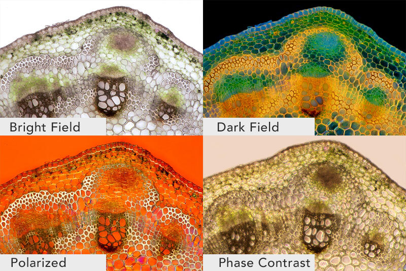
Four types of Light micrographs: Bright Field, Dark Field, Polarized, Phase Contrast. © Marek Mis/Science Source
The basic microscope we used as a kid is the standard “light microscope”. Simply put, it uses light and a set of lenses. The addition of filters, specialized mirrors, lasers, specific light spectrums, and other features gives us much more detail.
More advanced devices include Scanning Electron Microscopes (SEM), Transmission Electron Microscopes (TEM), Atomic Force Microscopes (AFM), and Scanning Tunneling Microscopes (STM).
Commonly used techniques when viewing slides on a light microscope are Bright Field, Dark Field, Fluorescence, Differential Interference Contrast (DIC), Phase Contrast, and Confocal microscopy.
Light MIcroscope Bright Field: the light source shines directly from underneath the specimen, creating a light-colored or bright area around it.
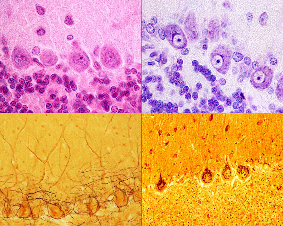
Purkinje neurons of the cerebellar cortex stained with four staining methods: hematoxylin eosin (top left), cresyl violet (top right), Cajal's silver nitrate (bottom left) and silver method for Golgi apparatus (bottom right). © Jose Luis Calvo/Science Source
Light Microscope Dark Field: the light source is occulted, so it reaches the specimen at different angles giving us slightly more varied details than if it was lit from directly underneath. The area around the specimen is dark or black.
Fluorescence: This uses light filters and specific wavelengths. Short wavelengths are reflected down to the specimen, which then fluoresces or gives off long wavelengths of light. These are reflected up to a mirror that allows long wavelengths to pass through to the lens.
Phase Contrast: Using a special lens and filters it allows viewing of transparent and colorless specimens. It looks similar to DIC micrography but lacks shadows, making it a bit more two dimensional.
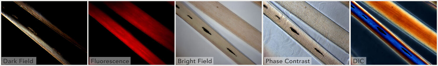
Different illumination techniques of a light microscope: dark field, fluorescence, bright field, phase contrast, DIC (differential interference contrast). Child’s hair strand © Ted Kinsman/Science Source
Differential Interference Contrast Microscopy (DIC): Using a polarizer, beam splitter, condenser, and filters it allows viewing of transparent and colorless specimens. It has a more three-dimensional appearance than phase-contrast microscopy.
Confocal Microscopy: Also called Confocal Laser Scanning Microscopy (CLSM), it uses a laser and a spatial pinhole to create a sharper image.
Let's look at the more advanced types of microscopes:

|Atomic Force Micrograph of plasma membrane proteins.
Scanning Electron Microscope SEM: Uses a particle beam of electrons. It detects reflected electrons off the surface of a specimen, which is placed in a vacuum. creating sharp images. Magnification ranges from 20x to 30,000x, spatial resolution of 50 - 100nm.
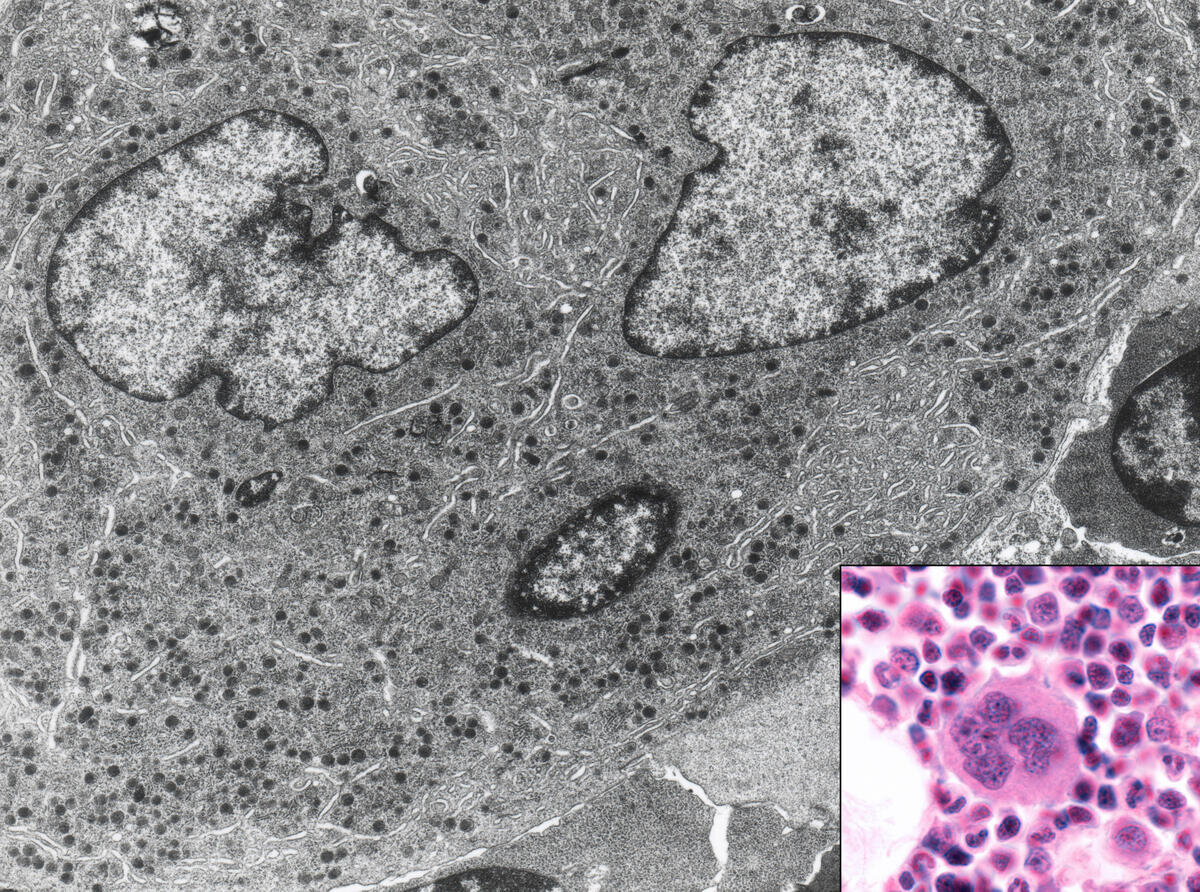
Bone marrow cell. This micrograph shows this cell with light (inset) and electron microscope. © Jose Luis Calvo/Science Source
Transmission Electron Microscope TEM: Uses a particle beam of electrons that pass through a thinly sliced specimen. It can show the internal structures of cells with a magnification up to 2,000,000x.
Atomic Force Microscope AFM: AFM uses a laser that bounces off of a stylus on a cantilever lever. This action traces the specimen. Any deviation triggers the sensors creating a raster image. One benefit of this is that it also records the Z-Plane. Another advantage of AFM over electron microscopy is that the specimen need not be in a vacuum.
Scanning Tunneling Microscope STM: Scanning Tunneling Microscope STM: An STM also uses electrons, based on quantum tunneling. The benefits are that it can be used in a vacuum, air, water, or ambient gas environment. It captures surfaces on the atomic level.
What can we look at with all of these scopes?
Scoop up pond water or ocean water to be astonished by the plethora of living zooplankton and phytoplankton visible within a single drop using a simple light microscope.
It opens you to the wonder of cyanobacteria, blue-green algae, ciliates like paramecium, daphnia, amoebas, and euglena. If you were lucky, you might have witnessed them conjugate and divide!
Additionally, the ocean water drop allows a peek at copepods, immature mollusks, krill, algae, crustaceans, fish in their zooplankton stage; and you may behold the breathtaking beauty of diatoms, the most common type of phytoplankton in our oceans.
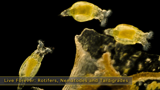
Switch to a higher-powered Scanning Electron Microscope (SEM) to view Water Bears, pollen, blood cells, and insects. An SEM uses a particle beam of electrons to photograph the surface of a vacuum-sealed specimen.
Transmission Electron Microscopes(TEM) allows us to see cross-sections of a specimen like the beautiful interior of the human body, marine life, and animal and plant cells. The TEM's particle beam passes through its vacuum sealed specimen.

Custom homedecor, phone cases, shirts and more.
Of course, there are critical medical applications.
We can view the cells of the human body with many different microscopes. The light mic, SEM, and TEM show scientists and medical researchers different angles and aspects of the cell and its fine structures and organelles.
Microscopes help scientists study cancer - breast, ovarian, prostate, liver, and skin cancer. We can develop an improved understanding of skin conditions such as psoriasis and eczema. They assist in the fight against nervous, respiratory, and circulatory system diseases. And a cure for muscular conditions such as fibromyalgia and multiple sclerosis (MS) and autoimmune conditions.
Infectious agents such as bacteria, viruses, fungi, prions, and parasites can be examined. It allows us to better understand, diagnose, and work towards cures, vaccines, or prevention.
And without the microscope, how could we progress in the fight against the seasonal flu, measles, polio, malaria, and HIV/AIDS.
If light, lasers, electrons, and quantum physics are not enough, there is even a microscopy method, similar to SONAR, that uses sound waves; Acoustic Microscopy.
Lastly, without these microscopes, we would not be able to continue our current fight against the Coronavirus, COVID-19.


No comments:
Post a Comment