Pages

Tuesday, January 5, 2021

Taking a Closer Look at Microscopy for Medical and Scientific Use

Waterbear Micrograph

 Read any scientific or medical news story and you can see that microscopy has come a long way since your high school biology class microscope.

It isn't just higher magnification, but crisp details, a greater depth of field, viewing internal features, and colorful 3D-like visuals that fascinate us. There have been many advancements to light microscopes and a multitude of new kinds that can see so much more than we could have ever expected.  

Stock Micrographs

Let's take a closer look!

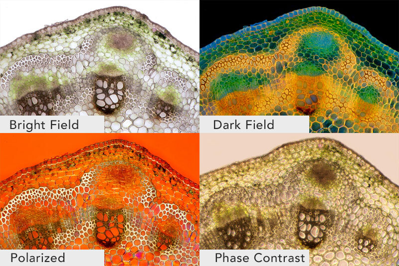
Four types of Light micrographs: Bright Field, Dark Field, Polarized, Phase Contrast. © Marek Mis/Science Source

Four types of Light micrographs: Bright Field, Dark Field, Polarized, Phase Contrast. © Marek Mis/Science Source

The basic microscope we used as a kid is the standard “light microscope”. Simply put, it uses light and a set of lenses. The addition of filters, specialized mirrors, lasers, specific light spectrums, and other features gives us much more detail.

More advanced devices include Scanning Electron Microscopes (SEM), Transmission Electron Microscopes (TEM), Atomic Force Microscopes (AFM), and Scanning Tunneling Microscopes (STM).

Commonly used techniques when viewing slides on a light microscope are Bright Field, Dark Field, Fluorescence, Differential Interference Contrast (DIC), Phase Contrast, and Confocal microscopy.

Light MIcroscope Bright Field: the light source shines directly from underneath the specimen, creating a light-colored or bright area around it.
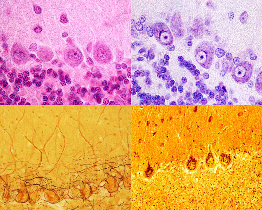
Purkinje neurons of the cerebellar cortex stained with four staining methods: hematoxylin eosin (top left), cresyl violet (top right), Cajal's silver nitrate (bottom left) and silver method for Golgi apparatus (bottom right). © Jose Luis Calvo/Science Source

Purkinje neurons of the cerebellar cortex stained with four staining methods: hematoxylin eosin (top left), cresyl violet (top right), Cajal's silver nitrate (bottom left) and silver method for Golgi apparatus (bottom right). © Jose Luis Calvo/Science Source

Light Microscope Dark Field: the light source is occulted, so it reaches the specimen at different angles giving us slightly more varied details than if it was lit from directly underneath. The area around the specimen is dark or black.

Fluorescence: This uses light filters and specific wavelengths. Short wavelengths are reflected down to the specimen, which then fluoresces or gives off long wavelengths of light. These are reflected up to a mirror that allows long wavelengths to pass through to the lens.

Phase Contrast: Using a special lens and filters it allows viewing of transparent and colorless specimens. It looks similar to DIC micrography but lacks shadows, making it a bit more two dimensional.

Different illumination techniques of a light microscope: dark field, fluorescence, bright field, phase contrast, DIC (differential interference contrast). Child’s hair strand © Ted Kinsman/Science Source

Different illumination techniques of a light microscope: dark field, fluorescence, bright field, phase contrast, DIC (differential interference contrast). Child’s hair strand © Ted Kinsman/Science Source

Differential Interference Contrast Microscopy (DIC): Using a  polarizer, beam splitter, condenser, and filters it allows viewing of transparent and colorless specimens. It has a more three-dimensional appearance than phase-contrast microscopy.

Confocal Microscopy: Also called Confocal Laser Scanning Microscopy (CLSM), it uses a laser and a spatial pinhole to create a sharper image.

Let's look at the more advanced types of microscopes:

|Atomic Force Micrograph of plasma membrane proteins.

|Atomic Force Micrograph of plasma membrane proteins.

Scanning Electron Microscope SEM: Uses a particle beam of electrons. It detects reflected electrons off the surface of a specimen, which is placed in a vacuum. creating sharp images. Magnification ranges from 20x to 30,000x, spatial resolution of 50 - 100nm.
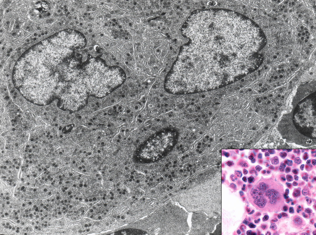
Bone marrow cell. This micrograph shows this cell with light (inset) and electron microscope. © Jose Luis Calvo/Science Source

Bone marrow cell. This micrograph shows this cell with light (inset) and electron microscope. © Jose Luis Calvo/Science Source

Transmission Electron Microscope TEM: Uses a particle beam of electrons that pass through a thinly sliced specimen. It can show the internal structures of cells with a magnification up to 2,000,000x.

Atomic Force Microscope AFM: AFM uses a laser that bounces off of a stylus on a cantilever lever. This action traces the specimen. Any deviation triggers the sensors creating a raster image. One benefit of this is that it also records the Z-Plane. Another advantage of AFM over electron microscopy is that the specimen need not be in a vacuum.

Scanning Tunneling Microscope STM: Scanning Tunneling Microscope STM: An STM also uses electrons, based on quantum tunneling. The benefits are that it can be used in a vacuum, air, water, or ambient gas environment. It captures surfaces on the atomic level.

What can we look at with all of these scopes? 

Scoop up pond water or ocean water to be astonished by the plethora of living zooplankton and phytoplankton visible within a single drop using a simple light microscope. 

It opens you to the wonder of cyanobacteria,  blue-green algae, ciliates like paramecium, daphnia, amoebas, and euglena. If you were lucky, you might have witnessed them conjugate and divide!

Additionally,  the ocean water drop allows a peek at copepods, immature mollusks, krill, algae, crustaceans, fish in their zooplankton stage; and you may behold the breathtaking beauty of diatoms, the most common type of phytoplankton in our oceans. 

Switch to a higher-powered Scanning Electron Microscope (SEM) to view Water Bears, pollen, blood cells, and insects. An SEM uses a particle beam of electrons to photograph the surface of a vacuum-sealed specimen.

Transmission Electron Microscopes(TEM) allows us to see cross-sections of a specimen like the beautiful interior of the human body, marine life, and animal and plant cells. The TEM's particle beam passes through its vacuum sealed specimen. 

micrograph-shower-curtain.png

Custom homedecor, phone cases, shirts and more.

Of course, there are critical medical applications. 

We can view the cells of the human body with many different microscopes. The light mic, SEM, and TEM show scientists and medical researchers different angles and aspects of the cell and its fine structures and organelles. 

Microscopes help scientists study cancer - breast, ovarian, prostate, liver, and skin cancer.  We can develop an improved understanding of skin conditions such as psoriasis and eczema. They assist in the fight against nervous, respiratory, and circulatory system diseases. And a cure for muscular conditions such as fibromyalgia and multiple sclerosis (MS) and autoimmune conditions.

Infectious agents such as bacteria, viruses, fungi, prions, and parasites can be examined. It allows us to better understand, diagnose, and work towards cures, vaccines, or prevention. 

And without the microscope, how could we progress in the fight against the seasonal flu, measles, polio, malaria, and HIV/AIDS.

If light, lasers, electrons, and quantum physics are not enough, there is even a microscopy method, similar to SONAR, that uses sound waves; Acoustic Microscopy.

Lastly, without these microscopes, we would not be able to continue our current fight against the Coronavirus, COVID-19.


Tuesday, December 22, 2020

Medical Micrographs: a View into the Human Body


science has advanced exponentially in the last century. Even just the way the human body and pathology are viewed. We’ve gone from the basic x-ray to be able to see a single cell in the human body. Or even smaller.

One of the most fascinating innovations has been the Scanning Electron Micrograph, often referred to simply as an SEM for short. It has revealed hidden worlds of the human body, medical, and of course the natural world as well.

Gallery of Medical and Anatomical SEMs (micrographs) Stock Photography

The scanning electron microscope was invented in 1937 by Manfred von Ardenne the SEM machine uses electrons to record the surface topography of objects.

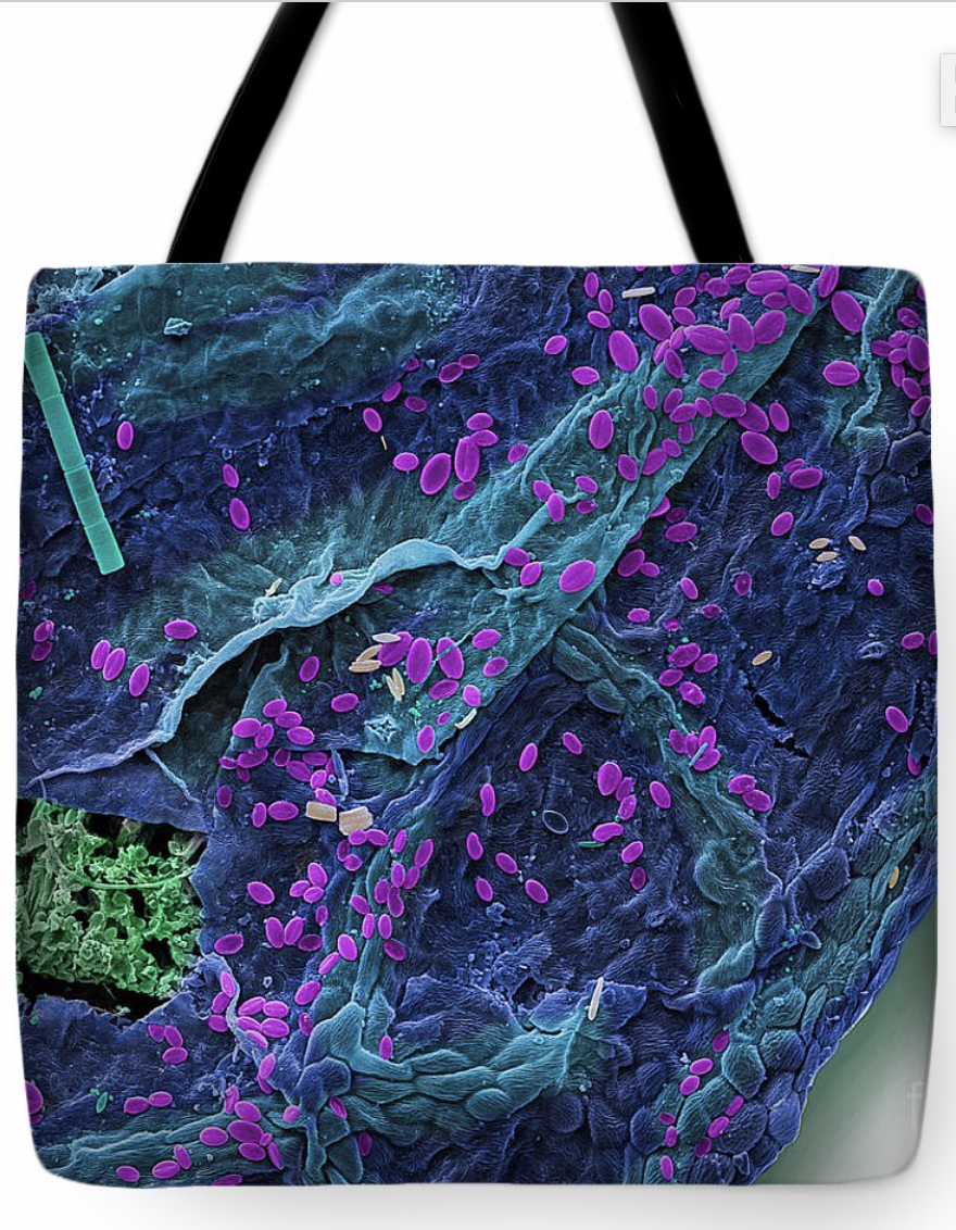
SEM of Maple Leaf Tote bag

SEM of Maple Leaf Tote bag

Thursday, February 13, 2020

Bringing to Light, Creatures of Darkness

Danté Fenolio, Ph.D., has devoted his life to bringing rarely seen creatures to light through his beautiful and eerie photography.
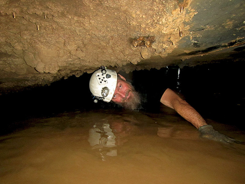
Danté Fenolio Caving in China
He's brought us images from habitats without light: caves, the bottom of the ocean, and the canopies of the rainforest. Not interested in the typical, but seeking out underappreciated and rarely seen creatures to highlight the need for conservation of the world's biodiversity.
Science Source Images is proud to represent several exclusive photographs from his collection. Discover a Fangtooth from the Gulf of Mexico, a colony of Siphonophore, orange and red-colored Jellies, transparent Batfish, blind albino Salamanders. Creatures so unusual, they may not seem real, but they are part of the wonder of our planet.
Fenolio is not only a successful author, adventure photographer, but runs the Conservation and Research Department at the San Antonio Zoo. His many projects to save animals and their habitats span the globe. Along with China, Peru, Japan, and Chile he works in the United States. His efforts range from the Ozarks, Oklahoma to Georgia.
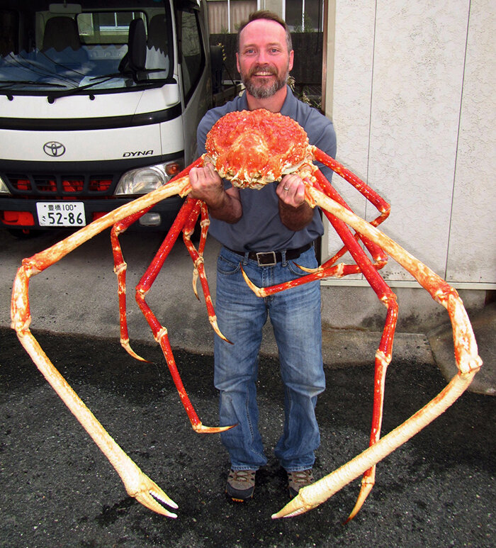
Bringing focus to blind cave fish, Japanese Giant Salamander and endangered Chilean amphibians.

Friday, December 20, 2019

The History of Christmas

an image of a blimp delivering christmas gifts, etching


As people put up Christmas trees and decorate their homes, it’s easy to believe these traditions have been around since the year one, but Christmas and the holidays surrounding December 25th have taken many forms.

The origin of Christmas comes from three sources: it is the date of the winter solstice on the Roman calendar, it is about nine months from the vernal equinox (March 25th) and it is the date set for the birth of Christ.

Before Christendom the Roman Empire celebrated Saturnalia, a holiday honoring the Roman god, Saturn, the farther of Jupiter. During Saturnalia social customs were flipped upside down. Slaves ate at their master’s tables, women flirted with men and gambling was permitted. Saturnalia also involved public banquets and gift giving, which may have influenced later Christian traditions. There were even reports of naked caroling!

Origins of Christmas stock Image Gallery

Cards, prints, tote-bags and more

The Christmas tree has its origins in 16th century Germany. While writing a sermon at night in a starry evergreen forest, the Protestant Christian reformer, Martin Luther, is said to have added lighted candles to a tree and put it into his house. Other Protestant Germans brought similar trees into their homes and the tradition spread. By the second half of the 19th century, the tradition had moved beyond Germany, first among the upper classes and eventually into lower areas of society.


While you are making your shopping rounds this holiday season, attending religious services or making time to be with family, reflect on the long history behind the season. Be sure to also check out great holiday gifts below and click the links above to see more historical images of the origins of Christmas.

From all of us from Science Source Merry Christmas and Happy Holidays!


Friday, November 1, 2019

Rising Sea Levels

One of the most significant challenges related to global climate change is rising sea levels. Since the beginning of the 20th-century, sea levels have gradually risen by 16 - 21 cm (6.3 - 8.3 in).

The rise in sea levels has also accelerated over time due to thermal expansion (the change in the volume and area of matter due to increased temperatures) and the melting of ice sheets and arctic glaciers.


Researchers have calculated that arctic sea ice has declined by 10% in the last 10 years. In addition to rising sea levels, this has altered the circulation of the Atlantic Ocean, destroying fisheries in northern countries and creating massive storms and hurricanes.

Coastlines around the world remain susceptible to flooding. Some at-risk coasts include the Miami shore, Rio de Janeiro, Osaka and Shanghai. The Egyptian city, Alexandria, faces a particular threat as much of its population exists in low lying coastal areas, which will likely be inundated within the next decade.


Science phone cases, mugs and more

Science phone cases, mugs and more
Our best hope for limiting the increase in sea levels is to reduce our carbon footprint. Initiatives in renewable energy and efficient power are critical components in reaching our emission goals.
For images of climate science and rising sea levels, click the links above. For information on how you can help deduce our carbon emissions head to the Environmental Defense Fund. (www.edf.org)

Thursday, September 26, 2019

Plastic is Deadly

Young Northern Fur Seal tangled in a nylon rope and dragging nets, cables, and fishing wires, doomed to die of hunger.
While plastic items in our daily lives are designed for one-time use, they have a long life span, usually up to 1,000 years.
Plastic is also deadly. People consume thousands of plastic articles each year, which damage our health, especially our immune systems. Plastic particles are even in the air we breathe, floating in the remotest places from fields to mountains.
For many animals plastic is lethal. Sea birds get caught in plastic fishing line and starve to death. Small mammals choke and die in 6-pack plastic rings. Dolphins and whales swallow plastic particles that block up their bodily systems. Sea turtles suffocate in plastic bags and floating debris prevents them from surfacing for air.
Luckily, as awareness grows, people are working to clean up plastic in the environment. New types of safe, biodegradable plastic are also coming on the market. We have a very long road ahead, but with more awareness and countries working towards a common goal, we have a more hopeful future.
plastic pollution, wildlife, animals, plastic in our lives

Marine Animal Custom Gifts: Framed Prints, Tote Bags, Note Books, and More…

Tuesday, August 6, 2019

Endangered Animals


Species around the world are facing extinction from threats ranging from ivory poaching to global climate change. Organizations such as the World Wildlife Fund have responded to the crisis with far-reaching conservation efforts.

The black rhinoceros has long been a victim of poaching for the international trade in rhino horn. British colonists began hunting rhinos in Africa during the colonial era and poaching has continued to this day. Conservationists work to protect rhino populations by building conservations and training local rangers to fend off poachers, but victories are hard fought. The black rhino’s population has sunk to 2,300 in recent years.

The polar bear has risen to public awareness due to the impact of climate change on the Arctic. Since 1979 the volume of sea ice shrunk by 80%, dramatically reducing the polar bear’s natural habitat. Experts say that the population of polar bears decreased to 20,000 to 25,000 in the last decade, classifying them a ‘vulnerable’ species. Conservationists work with businesses and governments to support green energy initiatives and help mitigate climate change.

Endangered Species Stock Image Gallery

It is worth noting that animals that enter the endangered species list do not always stay there. Famously, the American buffalo was reduced to near extinction following the American expansion into the western frontier but conservation efforts have increased their numbers gradually throughout the 20th century.

Science mugs, prints and cellphone cases

Likewise, the giant panda faced extinction a decade ago but has recently made gains due to protected habitats in China. Another encouraging story comes from the Siberian tiger, which was reduced to double digits in the early 1900s but has since increased to about 500.

While species around the world become endangered, conservationists continue to fight for their survival. Find out how you can help by visiting the World Wildlife Fund below. For a full gallery of endangered species follow the link and be sure to check out our storefront for great animal gifts.

The World Wildlife Fund

Science prints, tote bags and cellphone cases


  • wwf.org
  • rhinos.org
  • telegraph.com


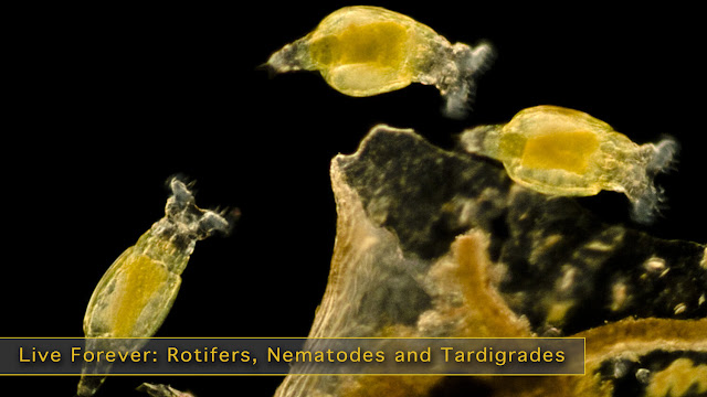
Rotifers, Nematodes and Tardigrades Stock Microscopic Photography

 Roti fers (Philodina sp.), Light Micrograph The bdelloid rotifer, found in freshwater habitats all over the world, is able to withstand ex...